Feline urticaria pigmentosa is a form of a condition known as cutaneous mastocytosis and it is caused by the accumulation of the defective mast cells (a type of white blood cells) in the skin, bone marrow, liver, spleen and lymph nodes. This skin condition is overall poorly documented, but it is better described in Sphynx and Devon Rex cats.
Background information
Mast cells, or mastocytes, are a type of white blood cells derived from the myeloid stem cells. Mastocytes contain granules rich in histamine and heparin and play important roles in allergic reactions, anaphylaxis, healing, angiogenesis, immune tolerance and blood-brain barrier function. In mastocytosis, mast cells accumulate in the skin and other tissues.
In humans, the most common type of mastocytosis is urticaria pigmentosa. This condition has also been described in dogs and cats, particularly Himalayan (Muller et al, 2001), Sphynx (Vitale et al, 1996) and Devon Rex cats (Nolie et al, 2009).

Feline urticaria pigmentosa
Causes
Genetic predisposition to urticaria pigmentosa has been identified in humans, but is also suggested in the Sphynx and Devon Rex cats. In humans, the majority of cases are caused by a point mutation in the proto-oncogene c-kit. This changes amino acid 816 in the c-kit protein. This is a transmembrane protein which binds to Mast Cell Growth Factor (MCGF) and signals the cell division. The respective mutation causes for the signal to be transmitted constantly causing abnormal cell proliferation and, ultimately, mastocytosis.
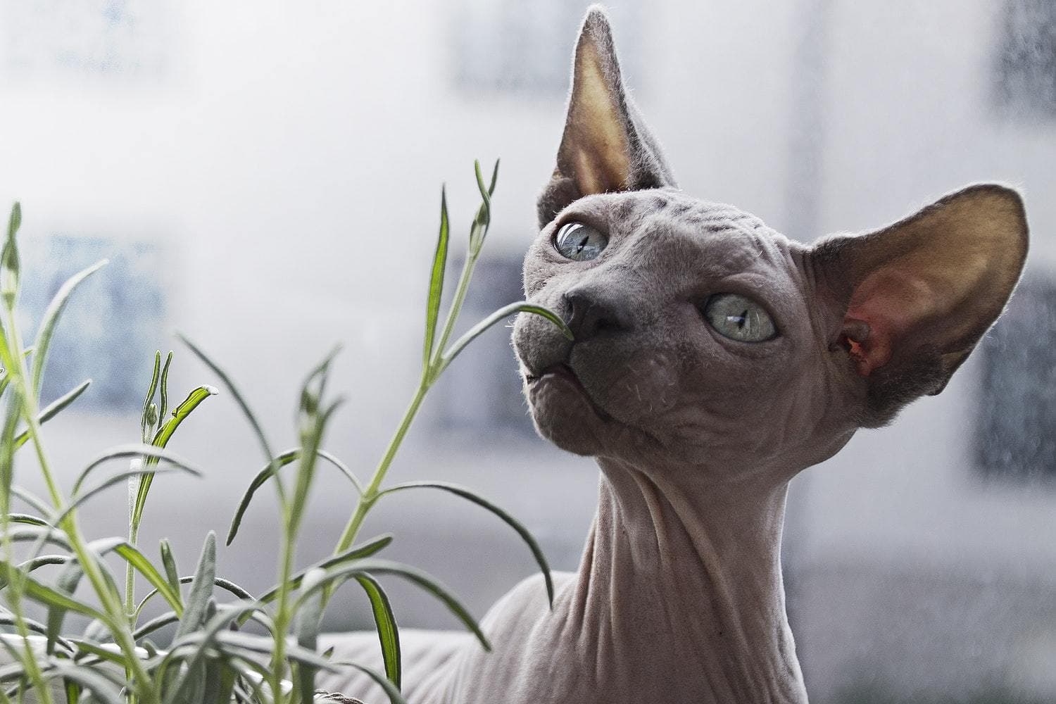
The direct cause of feline urticaria pigmentosa remain unknown. Because the condition has been primarily identified in naked cat breeds, studies suggest (Nolie et al, 2009) that the disease may be related to this phenotype. However, it can also be speculated that some cases of urticaria pigmentosa remain hidden and unidentified in fully covered cats.
Furthermore, both in humans and cats, it is supposed that this skin disease is closely related to allergic phenomenon which can trigger and/or worsen dermatitis. Irritants known to worsen the symptoms of feline urticaria pigmentosa are parasites, environmental allergens, food allergens etc.)

Feline urticaria pigmentosa
Symptoms and diagnosis
The clinical presentation of the disease is triggered at young age. The most common clinical signs are typical skin lesions – erythematous papules (red spots). Their distribution can be bilinearly symmetric and linear on the ventral lateral side of the trunk or diffuse on the ventral thorax. A case with greasy seborrhea on the head has been noted too (Nolie et al, 2009). Pruritus can be shown too. The clinical and histological presentation is similar between cases among Sphynx and Devon Rex cats. The diagnosis is achieved through skin biopsy and histopathological analysis.
Prognosis and therapy
The clinical prognosis of feline urticaria pigmentosa is generally good. However, this condition can be systematic and affect inner organs. Because it is so rare and poorly documented, it can be difficult to exclude this systemic progression. Human urticaria pigmentosa is treated symptomatically primarily based on avoidance of factors triggering the acute mediator release. Mast cell stabilizing agents, antihistamines and topical steroids can be applied too. In cats, the condition is generally successfully controlled with glucocorticoids, essential fatty acids (oral supplements) and anti-histamines.

Feline urticaria pigmentosa
Urticaria pigmentosa is a skin condition identified in Himalayan, Sphynx and Devon Rex cats. The documented cases all share similar clinical and histological presentation, but, unfortunately, there is still a lot we don't know about this rare skin condition.
Although generally successfully controlled, this condition does have a potential for systemic progression which is why it is important to be informed and keep an eye for clinical symptoms. This is particularly important for pet humans caring for naked cats who seem to be predisposed.
Further reading:
1. Muller GH, Kirk RW, Scott DW (2001). Small Animal Dermatology, 6th edn. Philadelphia: W.B. Saunders: 997–9. 6.
2. Nolie C, Colombo S, Abramo F, Scarampella F (2004). Papular eosinophilic/mastocytic dermatitis (feline urticaria pigmentosa) in Devon Rex cats: A distinct disease entity or a histopathological reaction pattern?. Veterinary Dermatology 15: 253-259
3. Vitale CB, Ihrke, PJ, Olivry T et al. (1996). Feline urticaria pigmentosa in three related Sphinx cats. Veterinary Dermatology 7: 227–33.